
Myiasis Disease: Etiology, Clinical Manifestations, Diagnostic Approaches, Conventional and Herbal Strategies for Effective Management
Livy-Elcon Emereonye
Abstract
Myiasis is a parasitic infestation of humans and animals caused by dipterous fly larvae that invade living or necrotic tissues. It remains a neglected tropical condition with high prevalence in developing regions, particularly sub-Saharan Africa where climatic conditions, poor sanitation, chronic wounds, and limited access to healthcare facilitate transmission. In Nigeria, multiple hospital-based studies reveal persistent incidence among children, rural dwellers, and patients with untreated ulcers. This article presents a comprehensive review of the causes, epidemiology, pathophysiology, clinical features, diagnosis, and conventional treatment of myiasis, while also examining scientifically supported herbal approaches useful as adjunct therapies. Emphasis is placed on Nigeria-specific trends and public health implications. Integrating conventional medicine with validated herbal interventions may enhance treatment accessibility and outcomes in resource-limited settings.
Keywords: Myiasis, fly larvae infestation, tropical parasitic diseases, ivermectin, herbal medicine, Nigeria, wound care
Introduction
I will like to state that myiasis is neither an STI nor STD. It is not transmitted through sexual contact, body fluids, or intimate relations like sexually transmitted infections/diseases.
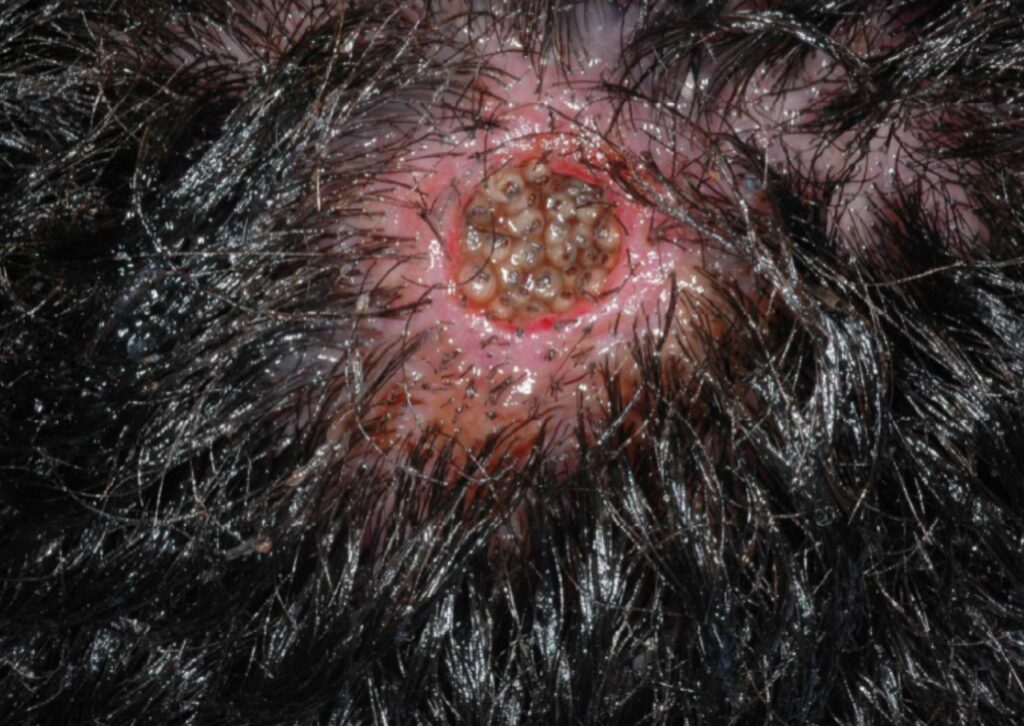
Myiasis is a parasitic disease caused by the invasion of human or animal tissue by larvae of certain dipterous flies. The larvae feed on living tissue, necrotic material, or bodily fluids, producing local tissue destruction and systemic complications when untreated. Although historically recognized in both medical and veterinary practice, myiasis continues to be underreported and neglected in public health frameworks, especially within tropical and subtropical regions.
The term myiasis was formally introduced by Hope in 1840, though systematic descriptions were later advanced by Zumpt in his classical monograph. While modern sanitation and healthcare have drastically reduced cases in industrialized nations, myiasis remains prevalent in Africa, Latin America, and parts of Asia. In Nigeria, environmental factors such as poor waste disposal, tropical humidity, open markets, proximity to livestock, and inadequate wound management contribute significantly to disease transmission.
Despite being largely preventable, myiasis continues to impose physical, psychological, and economic burdens on affected individuals. Chronic wounds complicated by larval infestation frequently progress to severe tissue necrosis, secondary infections, and social stigma. The disease is especially prevalent among vulnerable populations including children, elderly persons, homeless individuals, and patients with diabetes or peripheral vascular disease.
This article synthesizes current scientific knowledge on myiasis with a focus on Nigeria and tropical Africa, while highlighting emerging evidence on herbal therapies that may complement conventional management.
Epidemiology with Emphasis on Nigeria
Myiasis distribution correlates strongly with climatic conditions favoring fly breeding. Warm temperatures, high humidity, and inadequate sanitation create ideal environments for dipterous flies.
Sub-Saharan Africa reports a predominance of furuncular myiasis caused by Cordylobia anthropophaga, commonly known as the tumbu fly. This species deposits eggs on soil, clothing, or bedding, which hatch into larvae that penetrate human skin upon contact.
In Nigeria, several hospital-based and community studies indicate persistent myiasis occurrence:
Research conducted in southeastern Nigeria documented myiasis prevalence among primary school children ranging from 2% to over 7%, particularly during the rainy season when fly density peaks. Another clinical study in northern Nigeria reported wound myiasis in approximately 4–6% of patients presenting with chronic ulcers, diabetic foot wounds, and traumatic injuries.
In southwestern Nigeria, tertiary hospital reports have described recurrent cases of nasal and aural myiasis among elderly individuals with poor hygiene and untreated infections. Pediatric furuncular myiasis has been frequently associated with wearing un-ironed clothes dried outdoors, which harbor fly eggs.
Although comprehensive national surveillance data are lacking, these fragmented reports suggest that myiasis remains a persistent and underestimated parasitic problem in Nigeria, especially in rural and peri-urban communities.
Etiology and Causative Species
Human myiasis is caused primarily by larvae from several families of flies including Calliphoridae, Sarcophagidae, and Oestridae.
The most clinically significant species in Africa include:
– Cordylobia anthropophaga – the tumbu fly responsible for most furuncular myiasis cases
– Lucilia sericata – associated with wound myiasis
– Sarcophaga species – facultative wound invaders

Female flies lay eggs on decaying organic matter, wounds, or damp clothing. The larvae hatch within 24–72 hours and penetrate host tissues using mouth hooks and proteolytic enzymes.
Pathogenesis and Host Response
Once inside human tissue, larvae create a localized inflammatory reaction. They secrete enzymes that break down tissue proteins, allowing deeper penetration and feeding.
The pathological effects include:
– Mechanical tissue destruction
– Release of toxic metabolic products
– Secondary bacterial colonization
– Host inflammatory response
As larvae mature, they increase in size, intensifying pain and tissue damage. In wound myiasis, extensive necrosis may occur, occasionally exposing bone or deep structures.
If untreated, infestation can lead to systemic infection, tetanus, or in rare cases, septic shock.
Clinical Manifestations
Cutaneous (Furuncular) Myiasis
This is the most common presentation in Nigeria.
Patients typically develop:
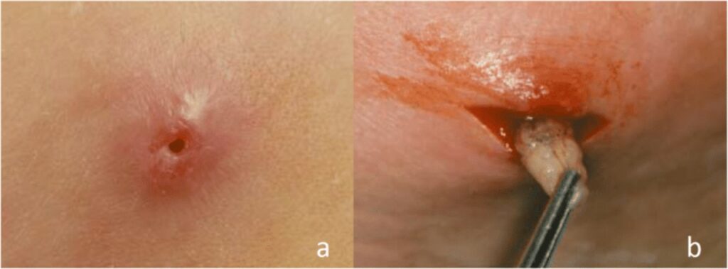
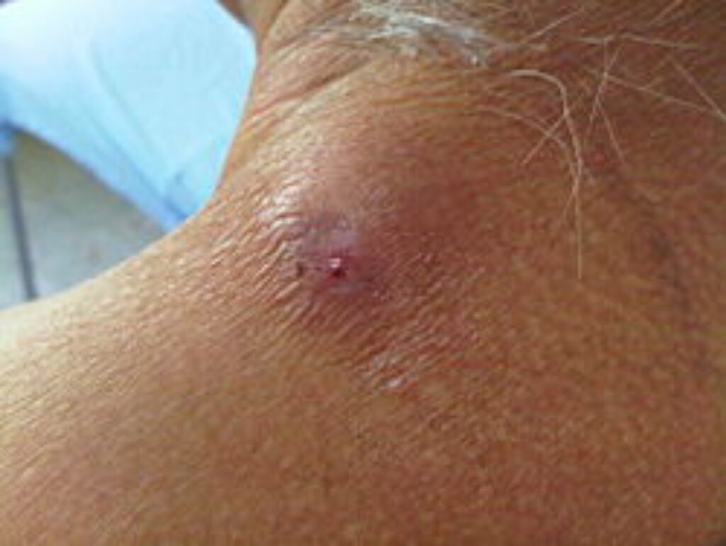
– Painful nodules resembling boils
– Central punctum through which larvae breathe
– Intermittent serous discharge
– Pruritus and burning sensation
– Sensation of movement beneath the skin
Lesions commonly occur on the trunk, buttocks, thighs, and scalp.
Wound Myiasis
Occurs in:
– Chronic ulcers
– Surgical wounds
– Traumatic injuries
Features include:
– Visible wriggling larvae
– Foul-smelling discharge
– Tissue necrosis
– Bleeding
This form is common among diabetic patients and individuals with neglected wounds.
Cavitary Myiasis
Involves body orifices such as:
– Nose (nasal myiasis)
– Ear (aural myiasis)
– Mouth and genitals
Symptoms include severe irritation, pain, bleeding, hearing loss, and facial swelling. In advanced cases, larvae may erode cartilage or bone.
Diagnostic Approach
Diagnosis is primarily clinical based on direct visualization of larvae.
Supportive investigations may include:
– Ultrasound imaging to locate subcutaneous larvae
– Endoscopic evaluation for nasal or aural cases
– Wound cultures for secondary infection
Laboratory identification of larvae is useful for epidemiological studies but not essential for routine treatment.
Conventional Medical Management
Mechanical Removal
The primary treatment involves careful extraction of larvae using forceps. Occlusive substances such as petroleum jelly or liquid paraffin are often applied to block oxygen supply, forcing larvae to surface.
Pharmacological Therapy
Ivermectin has demonstrated high efficacy in killing larvae and is especially useful in multiple infestations or inaccessible locations. A single oral dose of 200 micrograms per kilogram is often sufficient.
Antibiotics are prescribed when bacterial infection is present.
Tetanus prophylaxis is recommended in wound cases.
Wound Care
Debridement, saline irrigation, antiseptic dressing, and regular monitoring are crucial for healing.
Herbal and Traditional Approaches in Myiasis Management
Traditional medicine has played a significant role in parasitic disease management across Africa. Modern scientific research increasingly validates several herbal agents for larvicidal, antimicrobial, and wound-healing effects.
Neem (Azadirachta indica)
Neem extracts exhibit potent insecticidal and antimicrobial properties. Laboratory studies demonstrate that neem oil immobilizes and kills fly larvae by disrupting cellular respiration and membrane integrity.
In rural Nigeria, neem leaf decoctions are commonly applied to infested wounds to facilitate larval expulsion and prevent infection.
Turmeric (Curcuma longa)
Curcumin possesses strong anti-inflammatory and antimicrobial activities. It accelerates collagen formation and wound epithelialization, reducing healing time after larval removal.
Aloe vera
Aloe gel promotes fibroblast proliferation, reduces inflammation, and exhibits antibacterial effects. Its application after extraction soothes tissue irritation and enhances wound closure.
Eucalyptus Oil
Eucalyptus essential oil contains cineole, which demonstrates larvicidal and antiseptic effects. Studies confirm its ability to suffocate larvae while preventing microbial colonization.
Pawpaw (Carica papaya) Latex
Papaya latex contains papain enzymes that digest necrotic tissue and facilitate wound cleansing. It has been used traditionally to remove larvae and accelerate granulation tissue formation.
Integrative Therapeutic Perspective
Combining larval extraction, Ivermectin therapy, herbal wound-healing agents can provides a cost-effective strategy for rural healthcare settings. Such integration aligns with the World Health Organization’s endorsement of traditional medicine incorporation into primary healthcare systems.
Prevention and Public Health Implications
Myiasis is largely preventable through:
– Improved sanitation
– Fly control measures
– Public education on wound hygiene
– Proper clothing care (ironing dried clothes)
– Prompt medical attention for wounds
In Nigeria, strengthening waste management systems and primary healthcare outreach can significantly reduce disease incidence.
Conclusion
Myiasis remains a neglected yet preventable parasitic disease in Nigeria and other tropical regions. Environmental conditions, poor hygiene, and limited healthcare access perpetuate its persistence. Early diagnosis and rational treatment provide excellent outcomes. More research into herbal therapies with validated antimicrobial and larvicidal properties should be encouraged. Integrating scientific herbal medicine into standard care frameworks would improve accessibility and effectiveness of myiasis management.
References
Zumpt, F. Myiasis in Man and Animals in the Old World. Butterworths, 1965.
Francesconi, F., Lupi, O. Myiasis. Clinical Microbiology Reviews, 2012.
Ogo, N. I., et al. Human myiasis in Nigeria: clinical trends. African Journal of Clinical and Experimental Microbiology, 2020.
Akinbo, F. O., et al. Furuncular myiasis among school children in southern Nigeria. Nigerian Journal of Parasitology, 2014.
Robbins, K., et al. Human myiasis review. Journal of Infection in Developing Countries, 2014.
Nathan, S. S., et al. Larvicidal activity of neem oil. Bioresource Technology, 2005.
Gupta, S. C., et al. Curcumin therapeutic roles. Advances in Experimental Medicine and Biology, 2013.
Batish, D. R., et al. Eucalyptus oil larvicidal effects. Fitoterapia, 2008.
Cowan, M. M. Plant antimicrobials. Clinical Microbiology Reviews, 1999.
Leave a comment