
Lassa Fever: Etiology, Pathophysiology, and Treatment Approaches
By Livy-Elcon Emereonye
Abstract
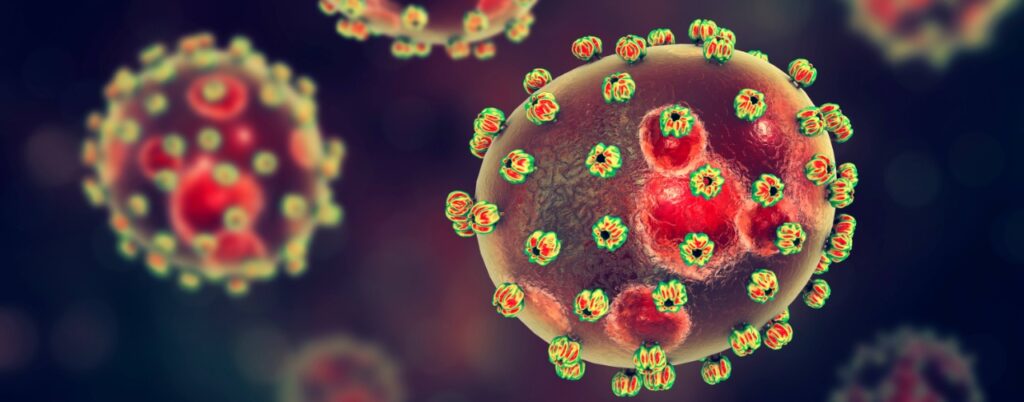
Lassa fever is an acute viral hemorrhagic illness endemic to West Africa and represents a major public health burden, particularly in Nigeria, Sierra Leone, Liberia, and Guinea. Caused by the Lassa virus, a member of the Arenaviridae family, the disease is primarily transmitted through exposure to excreta of infected Mastomys rodents and through person-to-person contact. The clinical spectrum ranges from mild febrile illness to severe multisystem disease characterized by hemorrhage, shock, and high mortality rates. This article comprehensively reviews the etiology, virology, pathophysiological mechanisms, clinical features, and current therapeutic approaches to Lassa fever, integrating emerging antiviral strategies and adjunctive supportive care modalities. Understanding the molecular and immunological mechanisms underlying disease progression is critical for improving diagnostic, therapeutic, and preventive strategies.
Keywords: Lassa fever, Arenavirus, viral hemorrhagic fever, ribavirin, pathophysiology, zoonotic disease
1. Introduction
Lassa fever is a zoonotic viral hemorrhagic disease first identified in 1969 in the town of Lassa located in Borno State, northeastern Nigeria. This historical link is why the disease was named Lassa fever. It is estimated that between 100,000 and 300,000 infections occur annually in West Africa, resulting in approximately 5,000 deaths. Nigeria experiences recurrent outbreaks with seasonal peaks during the dry season, posing significant challenges to healthcare systems.
Despite decades of research, Lassa fever remains underdiagnosed due to nonspecific early symptoms that resemble malaria, typhoid fever, and other febrile illnesses. The absence of a widely available vaccine and limited antiviral options further compound the disease burden.
2. Etiology and Virology
2.1 The Lassa Virus
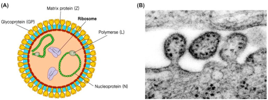
Lassa virus (LASV) is an enveloped, bi-segmented, single-stranded RNA virus belonging to the genus Mammarenavirus within the family Arenaviridae. The viral genome consists of:
– L segment encoding RNA-dependent RNA polymerase and Z protein
– S segment encoding nucleoprotein (NP) and glycoprotein precursor (GPC)
– The glycoproteins GP1 and GP2 facilitate viral entry via interaction with the α-dystroglycan receptor on host cells.
2.2 Reservoir and Transmission
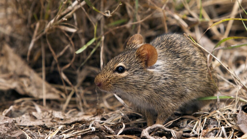
The primary reservoir is the multimammate rat (Mastomys natalensis), which sheds virus in urine and faeces throughout its lifetime.
Transmission occurs via:
– Inhalation of aerosolized rodent excreta
– Direct contact with contaminated food or surfaces
– Exposure to infected body fluids
– Nosocomial transmission
– Sexual transmission has been documented due to viral persistence in semen.
3. Epidemiology
Lassa fever is endemic in:
– Nigeria
– Sierra Leone
– Liberia
– Guinea
Nigeria reports thousands of confirmed cases annually, with case fatality rates ranging from 15–25% among hospitalized patients. Pregnant women in the third trimester exhibit mortality exceeding 30%, with foetal loss approaching 90%.
4. Pathophysiology
Lassa fever pathogenesis is multifactorial, involving viral replication, immune dysregulation, endothelial dysfunction, and organ damage.
4.1 Viral Entry and Dissemination
After mucosal or cutaneous entry, LASV infects:
– Macrophages
– Dendritic cells
– Endothelial cells
The virus spreads systemically through lymphatic and hematogenous routes.
4.2 Immune Evasion
LASV suppresses innate immune responses by:
– Inhibiting type I interferon production
– Blocking dendritic cell maturation
– Preventing effective T-cell activation
This results in uncontrolled viral replication.
4.3 Cytokine Dysregulation
Severe cases exhibit:
– Elevated TNF-α
– IL-6
– IL-8
Contributing to systemic inflammation and vascular leakage.
4.4 Vascular and Organ Damage
Infection of endothelial cells leads to:
– Increased permeability
– Plasma extravasation
– Hypovolemia
Major organs affected and pathology:
– Liver – Hepatocellular necrosis
– Kidney – Acute tubular necrosis
– Heart – Myocarditis
– Brain – Encephalopathy
– Inner ear – Sensorineural hearing loss
5. Clinical Manifestations
Early symptoms include:
– Fever
– Malaise
– Sore throat with exudates
– Headache
Progressive disease presents with:
– Vomiting
– Diarrhea
– Facial edema
– Hemorrhage
– Respiratory distress
– Permanent deafness occurs in up to one-third of survivors.
6. Diagnosis
Laboratory Methods:
– Reverse transcriptase-PCR (RT-PCR)
– ELISA for antigen and IgM antibodies
– Viral isolation (biosafety level 4 labs)
7. Treatment Strategies
7.1 Antiviral Therapy
Ribavirin remains the primary antiviral agent.
– Most effective when initiated within 6 days
– Reduces mortality by up to 90% in early disease
Mechanism includes inhibition of viral RNA synthesis and immune modulation.
7.2 Supportive Management
This includes:
– Aggressive fluid resuscitation
– Electrolyte correction
– Oxygen therapy
– Blood products
– Renal replacement therapy
7.3 Emerging Therapies include:
– Favipiravir
– Monoclonal antibodies
– Small-molecule viral entry inhibitors
– Vaccine candidates (DNA and recombinant viral vector platforms)
7.4 Herbal Approaches
Medicinal herbs like Garcinia kola, Andrographis paniculata, and Phyllanthus amarus exhibit strong antiviral properties. An evidence-baaed herbal remedy can be advantageous and also cost-effective.
7.5 Phytotherapeutic Immune Support as Adjunctive Therapy in Lassa Fever
The complex immunopathogenesis of Lassa fever involves early suppression of innate antiviral responses, impaired type I interferon signaling, dysregulated macrophage activation, endothelial dysfunction, and a hyperinflammatory cascade in severe disease. These mechanisms contribute to high viral loads, vascular leakage, multi-organ involvement, and susceptibility to secondary infections. While ribavirin remains the primary antiviral therapy when administered early, comprehensive supportive care aimed at immune modulation, infection control, and endothelial protection is essential for improving clinical outcomes. Within this context, phytotherapeutic immune-support formulations with broad-spectrum antimicrobial activity have attracted increasing scientific interest.
Infedi® is a natural immune-support formulation characterized by immunomodulatory, antiviral, antioxidant, and broad-spectrum antimicrobial properties, which may provide mechanistic relevance in the supportive management of Lassa fever. Bioactive phytochemicals commonly found in immune-enhancing botanicals—including flavonoids (such as quercetin and luteolin), phenolic acids, alkaloids, terpenoids, and saponins—have demonstrated antiviral activity against RNA viruses through inhibition of viral entry, suppression of viral RNA polymerase activity, interference with viral proteases, and modulation of intracellular signaling pathways such as NF-κB and MAPK. These actions contribute to reduced viral replication and enhanced expression of interferon-stimulated genes, thereby strengthening early innate immune defenses that are typically compromised in Lassa virus infection.
In addition to direct antiviral effects, immunomodulatory phytoconstituents enhance macrophage phagocytic capacity, stimulate natural killer (NK) cell cytotoxic activity, and promote balanced adaptive immune responses through optimized Th1/Th2 cytokine regulation and improved antigen presentation. Such immune enhancement may facilitate more effective viral clearance while preventing excessive inflammatory tissue damage. Furthermore, polyphenols, tannins, and essential oil components present in many medicinal plants exert broad-spectrum antimicrobial activity by disrupting microbial cell membranes, inhibiting enzymatic pathways, impairing biofilm formation, and interfering with quorum sensing mechanisms. This antimicrobial support is clinically relevant, as secondary bacterial infections frequently complicate severe viral hemorrhagic fevers and contribute to morbidity and mortality.
Oxidative stress plays a significant role in endothelial injury and vascular permeability during Lassa fever. Antioxidant phytochemicals within immune-support formulations scavenge reactive oxygen species, upregulate endogenous antioxidant enzymes such as superoxide dismutase and glutathione peroxidase, and stabilize endothelial function, thereby potentially mitigating hemorrhagic manifestations and organ damage. Through the combined effects of antiviral interference, immune system enhancement, anti-inflammatory regulation, antimicrobial protection, and oxidative stress reduction, adjunctive phytotherapeutic agents such as Infedi® present biologically plausible supportive strategies in Lassa fever care.
8. Prevention and Control should include:
– Rodent population control
– Food storage hygiene
– PPE use in healthcare settings
– Surveillance and rapid isolation
– Community education

9. Discussion
The hallmark of Lassa fever lies in immune suppression rather than overwhelming inflammation, distinguishing it from Ebola. The virus’s capacity to evade host defenses explains prolonged viremia and organ damage. Early ribavirin administration is critical but limited by late presentation in endemic regions.
Advancements in vaccine research and monoclonal antibody therapies offer hope for long-term disease control.

Herbal products that boost immunity and modulate the immune system will play a significant role in prevention.
10. Conclusion
Lassa fever remains a major zoonotic threat in West Africa, driven by environmental, socioeconomic, and healthcare infrastructure challenges. Its pathophysiology involves complex interactions between viral replication and host immune dysfunction. While ribavirin significantly improves outcomes, comprehensive prevention strategies and novel therapeutics are urgently needed.
Dr. Emereonye could be reached on: +234 803 3922 445
References (Verifiable)
1. World Health Organization. Lassa fever fact sheet. WHO, 2023.
2. McCormick JB et al. Clinical observations in Lassa fever. J Infect Dis. 1987.
3. Shaffer JG et al. Lassa fever in Nigeria. Emerg Infect Dis. 2014.
4. Richmond JK, Baglole DJ. Lassa fever epidemiology. Clin Infect Dis. 2003.
5. Yun NE, Walker DH. Pathogenesis of Lassa fever. Viruses. 2012.
6. Ogbu O et al. Lassa fever in West Africa. Lancet. 2007.
7. Khan SH et al. Ribavirin therapy in Lassa fever. J Infect Dis. 1999.
8. Bowen MD et al. Arenavirus molecular biology. Virology. 2000.
9. Fisher-Hoch SP et al. Effective ribavirin therapy. Lancet. 1995.
10. Sogoba N et al. Reservoir host dynamics. Am J Trop Med Hyg. 2012
Leave a comment